

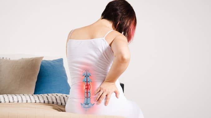
Do you feel pain in your tailbone, especially when sitting, but notice pain relief only when walking? If so, you might be dealing with coccydynia—commonly known as tailbone pain—which can often be alleviate with stretches for tailbone pain.
While it may not be severe, it can make everyday activities uncomfortable and frustrating.
Fortunately, specific exercises can help relieve the pain and improve your comfort.
1. Cobra
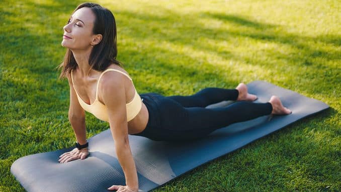
This stretch aims to open the chest and keep the back supple and healthy while regenerating the torso and relieving lower back pain.
- In a prone (faced down) lying down position, place the elbows on the floor under the shoulders with the hands forward.
- Stretch and lift the chest to the sky while extending the elbows and straightening the arms.
- Keep your hips in contact with the floor.
- Keep lifting out of the lower back.
- Squeeze the shoulder blades together.
- Squeeze the glutes while doing stretches for tailbone pain.
- Keep your gaze forward or slightly upward to avoid straining the neck.
- Breathe deeply as you hold this stretch for one minute.
If you do not have the flexibility to straighten the arms, lift as high as you can with the elbows bent.
2. Single Leg Knee Hug
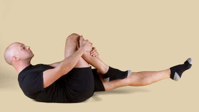
- Lie down on your back, then bend the left knee towards your chest until you feel a stretch.
- Hold for 30 seconds, then go back to the resting position.
- Repeat on the right knee.
- Perform each stretch 2–3 times per session, adjusting based on your comfort and mobility level.
This stretches for tailbone pain targets the piriformis and the iliopsoas muscles.
3. Figure 4 Stretch

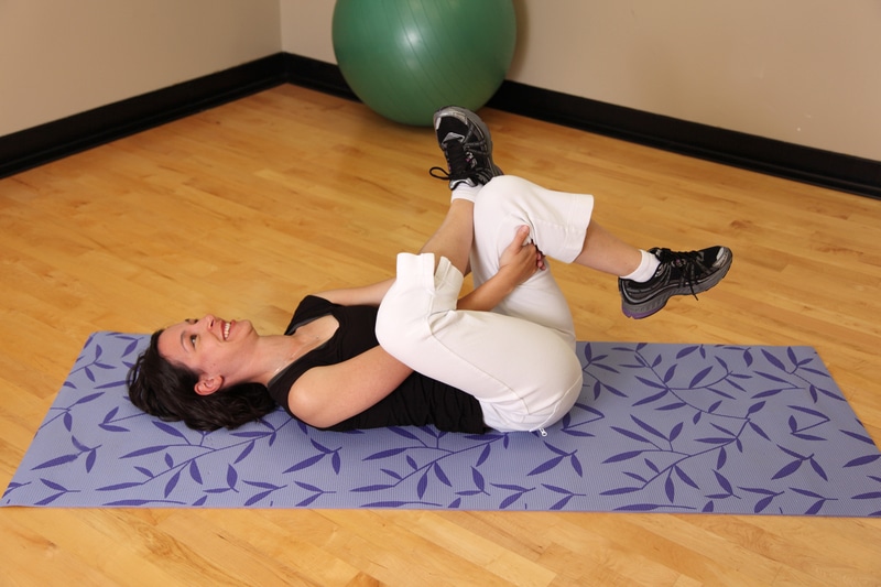
- Lie down on your back.
- Bend your knees towards your chest with your foot flat on the floor.
- Bend your left leg, put your left ankle across the right knee, and then hold your right thigh.
- Slowly pull it towards you until you feel a stretch.
- Ensure the foot of your non-stretching leg remains flat on the ground to maintain balance and alignment.
- Hold in for 30 seconds.
- Perform each stretch 2–3 times per session, adjusting based on your comfort and mobility level.
This stretches the piriformis as well as the gluteal muscles and is one of the effective stretches for tailbone pain.
4. Cat/Cow Stretch
This exercise aims to limber and stretch the back and neck muscles, stimulate spinal nerves, align the vertebrae, promote blood flow surrounding muscles, and relieve back and neck pain.
- Place the body in a prone (face-down) table-top position with the hands directly under the shoulders and the knees under the hips.
- Round your back up to the sky (Cat) and then arch your back with your head lifting up (Cow).
- As the Cat stretch is performe, exhale.
- As the Cat stretch is performe, inhale.
- Perform this exercise for one minute.
If the hand placement hurts the wrists, make the hands into fists to alleviate the pain.
5. Kneeling hip flexor stretch

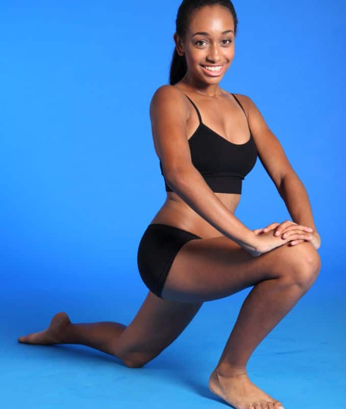
- Kneel on the floor, and bend your right leg forward at a 90-degree angle with your foot flat.
- Put a towel under your left knee for comfort.
- Rest your hands on your hips for stability.
- Tilt your pelvis anteriorly and slightly lean forward.
- Hold on for 30 seconds.
- Perform each stretch, 2–3 times per session, adjusting based on your comfort and mobility level.
This stretches the iliopsoas, which consists of the iliacus and then psoas muscles.
6. Kneel and twist
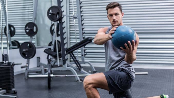
- Kneel on the floor, and bend your right leg forward at a 90-degree angle with your foot flat.
- Put a towel under your left knee for comfort.
- Then, raise your arms to shoulder height out to the side.
- Rotate your trunk toward the left side of the body and stop when the components are almost in line with the legs.
- Then slowly return to the resting position and do it on the right side.
- Do this for 7 repetitions, 1-2 sets.
This stretches the iliopsoas as well as improves mobility through the lower back.
7. Child’s Pose
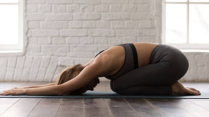
- Kneel on the floor while sitting on your knees.
- Then, place your hands flat on the floor.
- Slowly slide your body forward until you fully extend your arm while keeping your head down.
- Bring the forehead to the floor.
- Hold on for 30 seconds.
- Perform each stretch 2–3 times per session, adjusting based on your comfort and mobility level.
8. Reverse Kegels
The opposite of standard Kegels, this exercise focuses on relaxing rather than contracting the pelvic muscles. Reverse Kegels focus on releasing then relaxing the pelvic floor muscles.
This also helps remove an overly flexed tailbone, hypo-mobile and painful. Traditional Kegels concentrate on contracting and releasing the pelvic region. Both types can help balance your pelvic floor.
You can exercise while sitting, standing, or lying down on your back with your knees bent.
Once you’re in position, breathe deeply and bring awareness to your pelvic floor.
Feel your muscles relax and drop down as you inhale, incorporating stretches for tailbone pain.
- Find the position where you feel like sitting on your perineal tissue. This is the soft tissue between the anus and the scrotum in males or the anus and vagina in females.
- Place your hands on your belly and focus on taking diaphragmatic or belly breaths where, on the inhale, your belly expands.
- As your belly expands, focus on relaxing the muscles in your pelvic floor as you inhale, feeling the area gently expand and release tension.
- Keep that relaxed on the exhale and imagine further lengthening on the next inhale.
- With practice and improved body awareness, you should feel increased contact between your perineum and chair with a true reverse Kegel.
- Hold the reverse Kegel for 5 seconds and then release simultaneously.
- Do two to three sets of 10 throughout the day.
- Once you master this, you can try holding and releasing for extended periods.
Make sure you’re breathing while doing these exercises. It’s essential to live all the way into your belly as you inhale (instead of only breathing into your chest). Keeping your stomach relaxed helps.
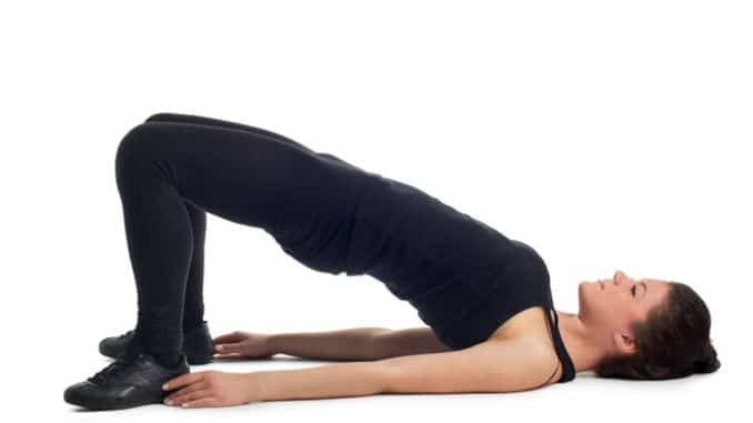
9. Pelvic Tilts
- Lie on your back with your knees bent and feet flat on the floor, maintaining good alignment with your head, shoulders, hips, and legs.
- Place your hands at your side with palms pressed on the floor.
- Tighten your abdominal muscles.
- Squeeze and tilt your pelvic floor muscles in and tilt your pelvis down pressing your low back into the floor.
- Hold this position for several deep belly breaths, in through your nose and out through your mouth.
- Relax and return to the starting position.
10. Hamstring Stretch
- Begin in an upright sitting position on the edge of a chair with your knees bent and feet flat on the floor.
- Maintain good alignment with your head, shoulders, and hips.
- Extend one leg forward with toes pointing towards the ceiling.
- Engage your core muscles.
- Hinge through your hips to move your upper body forward as you reach your hand towards your toes.
- Hold the position for several deep belly breaths through your nose and out through your mouth.
- Relax and repeat the movement on the opposite side.
11. Side-Lying Leg Lifts
- Lie on your side on the floor with your legs extended and stacked together.
- Bend your bottom arm to support your head.
- Maintain good alignment with your head, shoulders, hips, and legs.
- Tighten your abdominal muscles.
- Lift your top leg towards the ceiling.
- Hold the position for several deep belly breaths, in through your nose and out through your mouth.
- Relax and repeat the movement on the opposite side.
12. Butterfly Stretch
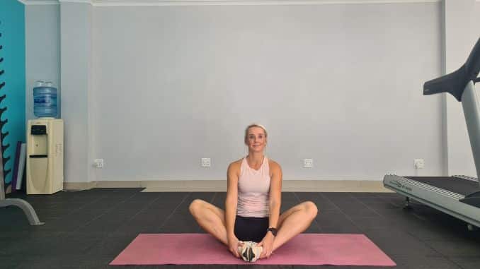
- Begin in an upright sitting position on the floor, maintaining good alignment with your head, shoulders, and hips.
- Engage your core and bring the soles of your feet together, spreading your knees out to the sides while keeping your spine straight.
- Hold the position for several deep belly breaths, in through your nose and out through your mouth.
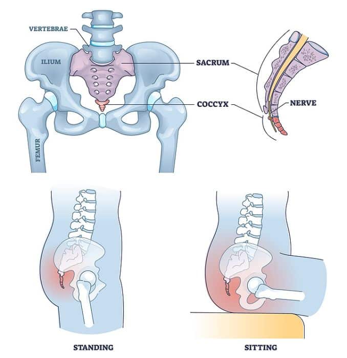
The Coccyx
The coccyx is a triangular-shaped bone located at the bottom of the spinal column, just below the sacrum.
It resembles a vestigial tail, thus the common name, tailbone, and can benefit from stretches for tailbone pain.
This bone can be damage or even fractured. This causes pain in your tailbone, worsening when sitting and improving when walking.
Using safe remedies at home and lifestyle modification may help manage this condition.

Usually, the coccyx has three to five bones fused or semi-fused together, depending on the person’s development.
The coccyx has limited mobility, but trauma, poor posture, or inflammation can result in pain and discomfort.
The coccyx joins the sacrum by the sacrococcygeal joints [3].
It is often neglect as it has no function and is only often vestigial or unnecessary in the human body.
The coccyx joins the sacrum by the sacrococcygeal joints.
However, it has a role in the pelvis.
When sitting, it supports the individual in a three-part support system.
It distributes the weight to provide balance and stability, helping with stretches for tailbone pain.
The lateral edges of the coccyx serve as a site for the insertion of the coccygeal muscles, the sacrospinous ligament, the sacrotuberous ligament, and the fibers of the gluteus maximus muscle.
Meanwhile, the iliococcygeus muscle tendon inserts onto the tip of the coccyx.
These different structures help and support the pelvic floor and contribute to defecation.
How does Coccydynia occur?
Trauma is the most common cause of this condition, such as falling directly on the coccyx or during childbirth.
Coccyx pathological instabilities are believe to be the root of inflammation and pain.
The pathological instability of the coccyx is said to be the culprit to pain and chronic inflammatory changes.
Obesity can also be a cause of coccydynia. The coccyx tends to stick out posteriorly when higher weight people sit down due to a lack of side pelvic rotation.
Because of this, there is increased exposure to intrapelvic pressure when sitting, which causes coccyx subluxation.
The coccygeal configuration types influence the pain level felt by an individual, and stretches for tailbone pain can help alleviate discomfort.
- Type I.- Coccyx is slightly curving forward, with its crown directed downward and extremely.
- Type II.- Apparent forward curvature of the coccyx and apex extends straight forward
- Type III.- The coccyx is more angulated forward
- Type IV.- The coccyx is subluxated.
From left to right, Type 1, Type 2, Type 3, and Type 4.
What are the Symptoms of Coccydynia?
The following symptoms characterize Coccydynia [1]:
- Localized pain and tenderness- Pain is localized in the area of the tailbone. No radiating pain is felt on the pelvis or the lower leg. Pain felt is described as aching soreness and can be mild or severe. There is constant tightness, pain, and discomfort around the area of the tailbone, mainly when pressure is applied.
- Worsen Pain when sitting- When sitting on a hard surface without a soft cushion or when a person leans backward in a sitting position, it puts extra pressure on the coccyx, causing pain to worsen or increase.
- Severe Pain when standing up from sitting- The movement from sitting to standing where the rotation of the pelvic bones and muscle movements that assist this rotation is painful.
Coccydynia Diagnosis
Coccydynia is often diagnosed through a thorough medical and physical examination.
The medical examination includes the current symptoms, how it was acquired or developed, lifestyle or if they had recent injuries, and more.
After the medical history is taken, a physical examination will be conducted.
It includes palpation, an Intrarectal exam, and manipulation.
During palpation, the doctor will palpate around the coccyx area to look for swelling and tenderness or even coccygeal spicules [4] (bone spurs), cysts, or tumors.
During intrarectal examination and manipulation, the doctor will manually try to manipulate the coccyx through the rectum to assess hypo or hypermobility of the sacrococcygeal joint.
It may also be used to examine or confirm if there is any muscle tension in the pelvis connecting to the coccyx.
Although not necessary, diagnostic procedures or tests can sometimes be done.
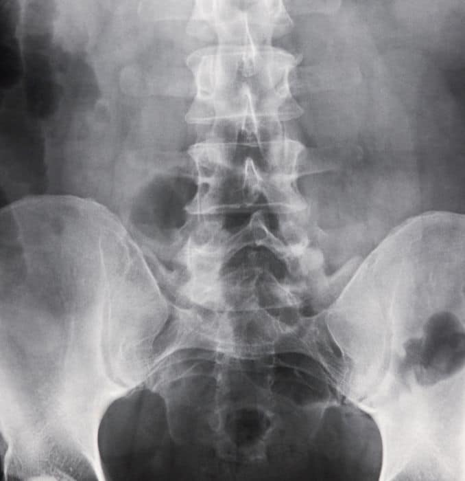
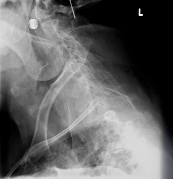
It includes dynamic X-ray imaging tests, Coccygeal discogram, computerized tomography, and Magnetic Resonance Imaging scans.
In a dynamic X-ray, two images will be produced to assess movements, such as those involved in stretches for tailbone pain.
One is when the patient is in a sitting position, and another is when the patient is in a standing position.
The doctor will then compare the two images and measure the angle of pelvic rotation and the coccyx’s position.
Indicating the condition is when the measurements are outside the normal range between 5 and 25 degrees.
 |  |

In a Coccygeal discogram, injection of local anesthesia on the sacrococcygeal region, either on the intervertebral joint or disc, is done.
It is to determine the exact location where the pain is coming from.
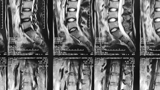
In Computerized Tomography (CT) and Magnetic Resonance Imaging (MRI), an image of the coccyx is produced to diagnose a fracture, tumor, or abnormal mobility of the sacrococcygeal joint.
In rare cases, a routine blood test is done to rule out infection or tumor.
Coccydynia Treatment
Dr. Melinda Avery, a specialist at the International Spine, Pain & Performance Center in Washington, D.C., emphasizes the importance of a multidisciplinary approach in managing coccydynia.
She notes that while conservative treatments like physical therapy, stretching, and manipulation have shown efficacy, there is a scarcity of high-quality studies supporting injection-based therapies such as corticosteroids and nerve blocks.
Surgical intervention, particularly coccygectomy, is considered for chronic or refractory cases, but more high-level comparative studies are needed to guide treatment decisions.
Most cases of coccydynia can be successfully treated with nonsurgical treatments and lifestyle modification. Treatments include:
- Non-steroidal anti-inflammatory drugs (NSAIDs) – ibuprofen (Advil), naproxen (Aleve), or COX-2 inhibitors (Celebrex) are examples of NSAIDs that can help reduce the inflammation and pain around the coccyx area.
- Cold Therapy/Ice Pack- Applying cold therapy or an ice pack to the area for the first 24-48 hrs of the injury can help reduce inflammation and pain.
- Heat Therapy/Heat Pads- Applying heat therapy or heat pads to the area can help reduce muscle spasms. You can also use a hot water bottle, chemical heat pack, long-lasting adhesive heat strip, or hot bath.
- Lifestyle Modification- Modifications to activities may help reduce pain, like using pillows, soft cushions such as U- or V-shaped pillows, and wedge-shaped pads with a cutout or hole where the tailbone is when sitting. It is to decrease the pressure on the coccyx.
- Dietary changes- Increased fiber and water intake are recommended if the pain increases during bowel movements, constipation, and stool softeners.
- Injection- Lidocaine, a numbing agent, and steroids are injected to decrease inflammation and pain in the coccyx area. The injection may last for a week up to several years, and they may receive up to 3 injections in a year.
- Manual Manipulation- Manual manipulation of the coccyx may be done to relieve pain. It involves adjusting the sacrum and coccyx joint to reduce pain.
- Massage- Spasms of the pelvic floor muscles can be massaged to alleviate pain. This causes added strain on the ligaments and sacrococcygeal joint, which limits mobility, thus pulling the coccyx.
- Stretching- Gentle stretches for the ligaments attached to the coccyx can help reduce muscle spasms.
If these nonsurgical treatments are ineffective, surgery to the coccyx may be recommended. This includes surgically removing all or a portion of the coccyx or coccygectomy.
Things to remember:
- Avoid sitting in a poor slumped posture, such as sacral sitting. Sitting this way decreases coccyx bone motion and can reinforce poor alignment.
- Avoid sitting longer than 15 to 20 minutes if you have coccyx persistent tailbone pain [2]. Shift your body weight as needed to decrease the tailbone pain, and if necessary, sit on a unique pillow called a donut to relieve tailbone pain.
- Consider consulting a pelvic floor therapist to learn techniques for releasing tension in this area safely.
- Avoid pushing with defecation and avoid constipation. Moving with bowel and bladder function can increase both coccyx and rectal pain.
Takeaway
Preventive measures may be taken, so you don’t acquire this condition.
Here some of the following:
- Use a Coccyx cushion – these can help reduce pressure on your tailbone when sitting.
- Limit prolonged sitting, stand or walk frequently, and use cushions designed for tailbone support.
- Avoid wearing tight jeans or trousers as it can put additional pressure on the tailbone.
Tailbone pain might be uncomfortable but is often temporary. Visit a doctor if symptoms become severe.
Do the necessary management and treatment to reduce tailbone pain, and it will recover quickly.
Unlock your best self with 11 Daily Stretches to Feel and Look Amazing. Improve flexibility, reduce stress, and boost confidence with simple yet effective moves. Start today and feel the difference in your body and mind.


Frequently Asked Questions (FAQs)
How do you self-adjust your back stretches for tailbone pain?
Try pelvic tilts, gentle stretches, or laying on your back with knees bent while rocking side to side.
However, if pelvic pain still persists, a chiropractor or physical therapist may help.
How do you sleep if your tailbone hurts?
Sleep on your side with a pillow between your knees or on your back with a cushion under your lower back and knees for support. Avoid sleeping directly on your tailbone.
Should I massage my tailbone?
Yes, gentle massage can help relieve tension and improve blood flow, but avoid applying too much pressure. If the pain worsens, consult a professional.
How do I know if my tailbone pain is serious?
If the pain is severe, lasts more than a few weeks, worsens over time, or is accompanied by numbness, swelling, or difficulty controlling bowel or bladder function, seek medical attention.
1. Published: 07 May 2008, Volume 1, pages 223–226, (2008)
Patel, R., Appannagari, A. & Whang, P.G. Coccydynia. Curr Rev Musculoskelet Med 1, 223–226 (2008). https://doi.org/10.1007/s12178-008-9028-1
2. Wilking, Ashley N. MS, PA-CAuthor Information
Journal of the American Academy of Physician Assistants 32(4):p 54-56, April 2019. | DOI: 10.1097/01.JAA.0000554227.56253.88
3. Perkins, Richard; Schofferman, Jerome; Reynolds, JamesAuthor Information
Journal of Spinal Disorders & Techniques 16(1):p 100-103, February 2003.
4. Original Article, Published: 06 January 2015, Volume 24, pages 1102–1108, (2015)
Doursounian, L., Maigne, JY. & Jacquot, F. Coccygectomy for coccygeal spicule: a study of 33 cases. Eur Spine J 24, 1102–1108 (2015). https://doi.org/10.1007/s00586-014-3753-5