 copy")
The anatomy of the knee includes the tibia. It is the largest bone in the body, aside from the femur. The talus, which forms the lower portion of the ankle, connects to the tibia’s lower extremity. The tibia’s upper end, which is located on the outer aspect of the leg, connects the tibia and femur. The fibula, unlike the tibia, does not bear weight. Instead, it contributes to the lateral aspect of the ankle as an ankle joint stabilizer and an attachment location for one of the four major knee ligaments and the biceps femoris tendon.
Anatomy of the Knee cont.
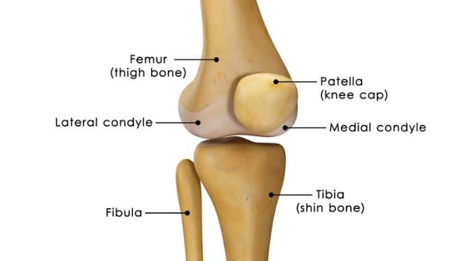
Patella
The patella, which is also known as the kneecap, protects the knee joint. It holds the quadriceps tendon on the lower end of the femur, acting as a fulcrum for the quadriceps muscles. A quadriceps is a group of four individual muscles on the anterior part of the thigh. The lower patella connects to the tibia through the patellar tendon.
Menisci
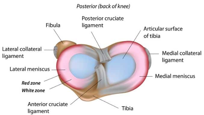
The medial and lateral menisci, known as C-shaped fibrocartilages, incompletely cover the surface of the tibia that joins with the femur. The menisci function as shock absorbers that equally spread the weight of the body, reducing friction between the tibia, and the femur during knee movements. They assist in knee rotation and play a function in stabilizing the ligaments.
Most areas of the menisci do not have a direct blood supply, which means they do not get the oxygen and nutrients needed for healing from the bloodstream. Damaged structures are difficult to repair. The menisci deteriorate with age. Menisci damage is a common cause of knee pain and injuries.
Knee Joint Capsule
The knee capsule is a thick, water-tight sac that surrounds the knee joint. The outer part of the capsule is lined with a fibrous and thick membrane. Within the capsule is the synovial membrane, which produces the fluid that lubricates the joints. This fluid also provides nutrients to the articular cartilage.
Muscles
The quadriceps and the hamstrings support the movements and stabilization of the knee joint. The quadriceps is actually composed of four individual muscles located on the anterior upper leg. These muscles are the vastus lateralis, vastus medialis, vastus intermedius and rectus femoris. These muscles fuse, forming the quadriceps tendon. The quadriceps straightens the knee by pulling the patella upon contraction.
The hamstrings are the muscles that attach to the tibia, specifically at the back of the knee. It consists of three individual muscles: biceps femoris, semitendinosus, and semimembranosus. The hamstrings functions by flexing or bending the knee joint. This muscle group also provides stability on both sides of the knee.
That is the end of Part 2. And I will have part 3 up very soon.
If you are interested in knee pain, knee injuries, or ACL injuries, these other posts may interest you:
- ACL Injuries in Female Athletes
- 3 ACL Exercise Mistakes
- What to do about Knee Pain?
- To Leg Press or Squat if You Have an ACL Injury?
- NCAA and ACL Injuries
- Downhill Walking Good After ACL Surgery?
- Knee Injuries and Exercises with Kevin Yates
If you are looking for stuff with more details on the anatomy of knee pain and ACL injuries, you can attend the live course called Exercise Rehabilitation of the Knee or instantly download my video presentation on Exercises for Prevention, Rehabilitation, and Overcoming Knee Injuries.
Rick Kaselj, MS